
Abstract
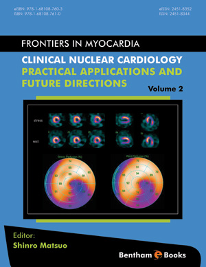
With the increasing availability of positron emission tomography (PET) myocardial perfusion imaging (MPI), PET MPI and the absolute quantification of myocardial blood flow (MBF) have become popular in clinical settings [1]. PET MPI shows higher diagnostic accuracy than that of single-photon emission computed tomography (SPECT) and shows predictive value for cardiac events [2, 3]. Quantitative MBF assessment also provides important additional diagnostic or prognostic information over that attained through conventional visual assessment [4]. The success of MBF quantification using PET/computed tomography (CT) has increased demand for this quantitative diagnostic approach to be more accessible. In this regard, MBF quantification approaches have been developed using several other diagnostic imaging modalities including SPECT, dynamic CT perfusion imaging, and cardiac magnetic resonance (CMR). In the United States (US), the Food and Drug Administration (FDA) has approved 13N-ammonia (13N-NH3) and 82rubidium (82Rb) for clinical use [5]. The Japanese Ministry of Health, Labour and Welfare (JMHLW) approved 13N-NH3 PET MPI for diagnosis of coronary artery disease (CAD) in March 2012 but has not approved other PET MPI tracers [6, 7]. Since 13N-NH3 PET MPI will be addressed elsewhere in this e-book, this review will address the clinical aspects of PET/CT MPI using other PET flow tracers.
Keywords: Blood Flow, Coronary Artery Disease, Endothelial Function, Flow Reserve, Positron Emission Tomography, Quantification.
Related Journals

Anti-Inflammatory & Anti-Allergy Agents in Medicinal Chemistry
Current Respiratory Medicine Reviews
Infectious Disorders - Drug Targets

Current Aging Science

Anti-Infective Agents
New Emirates Medical Journal
Current Psychopharmacology
Current Dentistry

Adolescent Psychiatry

Current Diabetes Reviews
Related Books
Andrology: Current and Future Developments
Male Infertility: An Integrative Manual of Western and Chinese Medicine

Awake Thoracic Surgery
The Anatomical Foundations of Regional Anesthesia and Acute Pain Medicine Macroanatomy Microanatomy Sonoanatomy Functional anatomy

Frontiers in Anti-infective Agents
Frontiers in Anti-infective Agents

Frontiers in Anti-infective Agents

Frontiers in Anti-infective Agents

Frontiers in Anti-infective Agents

Frontiers in Anti-infective Agents