
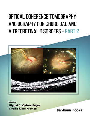
Abstract
Venous drainage from the retina merges into the central retinal vein and can be obstructed in the branch veins that drain the retinal quadrants, or the central retinal vein itself, which are termed Branch Retinal Vein Occlusion (BRVO) and Central Retinal Vein Occlusion (CRVO), respectively. Obstruction of retinal venous drainage often leads to a sudden or progressive increase in distal venous and capillary pressure with loss of vision and visual field defects. The extent of visual impairment correlates with the location and severity of the venous occlusion and how it impacts perfusion in the retina. Macular edema or retinal ischemia secondary to retinal vein occlusion is responsible for vision loss in retinal vein occlusions, and the advent of anti-VEGF therapeutics has revolutionized the management of vascular disease in the retina.
In this chapter, we review our current understanding of retinal vein occlusions and how OCT-Angiography (OCT-A) is being used clinically in the diagnosis and management of obstructive venous vascular phenomena. The benefits of using OCT-A in the diagnosis and management of CRVO and BRVO over conventional approaches, such as Fundus Fluorescein Angiography (FFA), are discussed. The current limitations of OCT-A and recent advances in the technology are also covered here. Finally, we assess how OCT-A can play a role in the development of new therapeutics to tackle one of the major causes of vision loss worldwide.
Keywords: Binarized, Branch retinal vein occlusion, Central retinal vein occlusion, Deep capillary plexus, Hemiretinal vein occlusion, Optical coherence tomography angiography, Macular edema, Neovascularization, Relative afferent pupillary defect, Retinal ischemia, Retinal vein occlusion, Skeletonization, Superficial capillary plexus.
Related Journals

Anti-Inflammatory & Anti-Allergy Agents in Medicinal Chemistry

Current Diabetes Reviews
Current Neurovascular Research
Current Respiratory Medicine Reviews
Current Pediatric Reviews
Infectious Disorders - Drug Targets
Current Stem Cell Research & Therapy
Endocrine, Metabolic & Immune Disorders - Drug Targets
Current Aging Science
Anti-Infective Agents
Related Books
Andrology: Current and Future Developments
Male Infertility: An Integrative Manual of Western and Chinese Medicine

Awake Thoracic Surgery
The Anatomical Foundations of Regional Anesthesia and Acute Pain Medicine Macroanatomy Microanatomy Sonoanatomy Functional anatomy
Frontiers in Anti-infective Agents
Frontiers in Anti-infective Agents

Frontiers in Anti-infective Agents
Frontiers in Anti-infective Agents

Frontiers in Anti-infective Agents

Frontiers in Anti-infective Agents