
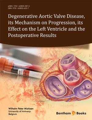
Abstract
The evaluation of the aortic valve can be performed in several ways.
The main imaging techniques for heart valves are:
- Doppler-echocardiography.
- Catheter based investigation.
- Computer tomography.
- MRI-MRA.
Each of these techniques has its advantages and drawbacks.
Trans thoracic echocardiography or TTE remains the standard imaging technique. It is widely used due to its availability, low cost, non-invasive character and low radiation burden. Several modalities exists. Drawbacks of the technique are the operator dependency, difficult imaging in patients with obesity and in cases with extensive calcification. Trans-esophageal echocardiography or TEE might offer better imaging, but the technique is invasive.
The description of the valve, and calculation of hemodynamic parameters such as jet velocity, mean TVG and AVA are the important parameters to estimate the severity of CAVS. For some of thes parameters, the continuity equation can be used. These parameters allow the categorization of the severity of CAVS. Each of these parameter has its advantages and drawbacks. Flow dependency is an important issue to deal with.
In asymptomatic patients, it is useful to assess the rate of progress of CAVS. This can give an indication when AVR for a given patien might become necessary. Assessment of the LVF and LVM are also important parameters to take into account.
Catheter examination has the advantage of direct measurement of TVG. The technique is invasive and can be recommended when echocardiographic data are inconclusive or are in conflict with clinical data. Usually, there is good correlation with echocardiographic data. If the presence of CAD is suspected, coronarygraphy should be performed.
CT and CMR are valuable adjuncts in the assessment of AVA. CT also allows the scoring of the calcium load, which is an important indicator for the severity of CAVS. CMR allows the flow mapping and imaging of the LV.
Keywords: Aortic valve area, arterio-valvular impedance, calcification, cardio CT, cardio MR, catheterization, Doppler duplex, echocardiography, jet velocity, left ventricular diameter, left ventricular function, left ventricular mass, low flow low gradient, transvalvular gradient.
Related Journals

Anti-Inflammatory & Anti-Allergy Agents in Medicinal Chemistry

Current Diabetes Reviews
Current Neurovascular Research
Current Respiratory Medicine Reviews
Current Pediatric Reviews

Infectious Disorders - Drug Targets

Current Stem Cell Research & Therapy

Endocrine, Metabolic & Immune Disorders - Drug Targets

Current Alzheimer Research

Current Aging Science
Related Books

Andrology: Current and Future Developments
Male Infertility: An Integrative Manual of Western and Chinese Medicine

Awake Thoracic Surgery
The Anatomical Foundations of Regional Anesthesia and Acute Pain Medicine Macroanatomy Microanatomy Sonoanatomy Functional anatomy

Frontiers in Anti-infective Agents
Frontiers in Anti-infective Agents

Frontiers in Anti-infective Agents

Frontiers in Anti-infective Agents

Frontiers in Anti-infective Agents

Frontiers in Anti-infective Agents