
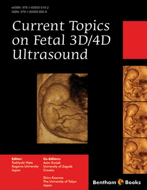
Abstract
We present herein the latest three-dimensional (3D)/four-dimensional (4D) sonographic studies on fetal heart and blood vessels using STIC (spatiotemporal image correlation) or real-time 3D fetal echocardiography. These new 3D/4D ultrasound techniques include VOCAL (Virtual Organ Computer-aided AnaLysis), TUI (Tomographic Ultrasound Imaging), SonoVCAD (Sonography Based Volume Computer Aided Diagnosis), color/power Doppler ultrasound and HD (High-Definition)-flow, B-flow and inversion mode. Fetal 3D/4D echocardiography provides a novel means for evaluation of fetal heart and blood vessels in 3D in real time, and of fetal intracardiac hemodynamics in the second and third trimesters. These novel techniques may assist in the evaluation of fetal cardiac anatomy and hemodynamics, and offer the potential advantages relative to conventional two-dimensional fetal echocardiography and Doppler flow mapping. 3D/4D ultrasound may be an important modality in future fetal cardiac research and in evaluation of congenital heart disease in the fetus.
Related Journals

Anti-Inflammatory & Anti-Allergy Agents in Medicinal Chemistry

Current Diabetes Reviews
Current Neurovascular Research

Current Respiratory Medicine Reviews

Current Pediatric Reviews
Infectious Disorders - Drug Targets

Current Stem Cell Research & Therapy
Endocrine, Metabolic & Immune Disorders - Drug Targets

Current Alzheimer Research

Current Aging Science
Related Books
Andrology: Current and Future Developments
Male Infertility: An Integrative Manual of Western and Chinese Medicine

Awake Thoracic Surgery
The Anatomical Foundations of Regional Anesthesia and Acute Pain Medicine Macroanatomy Microanatomy Sonoanatomy Functional anatomy

Frontiers in Anti-infective Agents
Frontiers in Anti-infective Agents

Frontiers in Anti-infective Agents

Frontiers in Anti-infective Agents

Frontiers in Anti-infective Agents

Frontiers in Anti-infective Agents